If your cervical screening test results suggest that you have a higher risk of developing cervical cancer, you will usually have more tests. You may also have tests to check for cervical cancer if you have symptoms.
Learn more about:
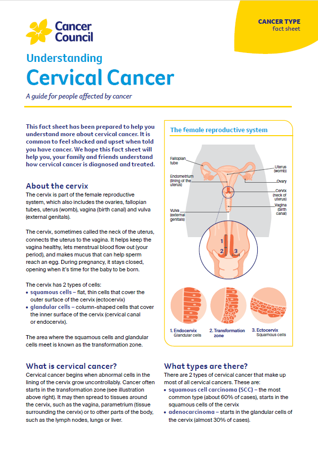
Overview
Some tests allow your doctor to see the tissue in your cervix and surrounding areas more clearly. Other tests tell the doctor about your general health and whether the cancer has spread. You probably won’t need to have all the tests described in this section.
If you feel anxious while waiting for test results, it may help to talk to a friend or family member, or call Cancer Council 13 11 20 for support.
Colposcopy
This looks closely at the cervix and vagina to see if there are any abnormal or changed cells. An instrument called a colposcope (a microscope with a light) is placed near your genital area but does not enter your body. An instrument called a speculum will be inserted into the vagina to spread the walls apart so the vagina and cervix can be seen more clearly.
Biopsy
If your doctor sees abnormal or changed cells during a colposcopy, they will usually take a small tissue sample (biopsy) from the cervix. The tissue sample will be sent to a laboratory to be examined under a microscope by a doctor called a pathologist. This can show if there are cancerous or precancerous cells.
Cone biopsy
This is done when a larger area of tissue needs to be removed or when early-stage cancer is suspected. A cone biopsy is usually done as day surgery in hospital under general anaesthetic. A surgical knife (scalpel) is used to remove a cone-shaped piece of tissue from the cervix. As with a biopsy (see above), the sample will be sent to a laboratory for testing.
I had period-like pain for a few days after the cone biopsy but a hot water bottle and mild pain medicines helped a lot.
Julie
Imaging scans
These can create pictures of the inside of your body and provide different types of information. You may have one or more imaging scans to check if the cancer has spread to other parts of your body. These scans may include an MRI, PET–CT scan or chest x-ray. To find out more about these scans, visit your local Cancer Council website or call Cancer Council 13 11 20.
A CT (computerised tomography) scan uses x-rays to take pictures of the inside of your body and then compiles them into a detailed, 3-dimensional picture.
When you make the appointment for the scan, you will be told if there are any special instructions to follow. Before the scan, you may be given a drink, or have an injection of a dye (called contrast) into one of your veins. The contrast may make you feel hot all over for a few minutes. You may also be asked to insert a tampon into your vagina. The dye and the tampon make the pictures clearer and easier to read.
During the scan, you will need to lie still on a treatment table that moves in and out of the CT scanner, which is large and round like a doughnut. The scan is painless and takes 5–10 minutes.
Learn more about CT scans.
An MRI (magnetic resonance imaging) scan uses a powerful magnet and radio waves to create detailed cross-sectional pictures of the inside of your body. Let your medical team know if you have a pacemaker or any other metal implant, as some may affect how an MRI works.
Sometimes gel is placed in the vagina before the MRI scan to better show the cervix or vagina. During the scan, you will lie on a treatment table that slides into a large metal cylinder that is open at both ends.
The test is painless but the noisy, narrow machine can make some people feel anxious or claustrophobic. If you are concerned, talk to your medical team before the scan. You may be given medicine to help you relax, and you will usually be offered headphones or earplugs. Most MRI scans take between 30 and 90 minutes.
Learn more about MRI scans.
A PET (positron emission tomography) scan combined with a CT scan is a specialised imaging test. It provides more detailed information about the cancer than a CT scan on its own. Not all people need to have a PET–CT scan.
Before having the scan, you will be injected with a glucose (sugar) solution containing a small amount of radioactive material. Cancer cells show up brighter on the scan because they take up more glucose than normal cells do. The radioactive material will leave your body within a few hours.
You will be asked to lie still for 30–60 minutes while the solution spreads through your body, then you will have the scan. Let your doctor know if you are claustrophobic, as you need to be in a confined space for the scan. It may take a few hours to prepare for a PET–CT scan, but the scan itself usually takes about 30 minutes.
Before having scans, tell the doctor if you have any allergies or have had a reaction to contrast during previous scans. You should also let them know if you have diabetes or kidney disease, or are pregnant or breastfeeding.
Examination under anaesthetic
To check whether the cancer has spread, the doctor may give you a general anaesthetic to examine your cervix, vagina, uterus, bladder and rectum. This is done in hospital and you can usually go home on the same day.
→ READ MORE: Staging for cervical cancer
Podcast: Tests and Cancer
Listen to more of our podcast for people affected by cancer
More resources

Dr Antonia Jones, Gynaecological Oncologist, Royal Women’s Hospital and Mercy Hospital for Women, Melbourne, VIC; Angelyn Aligarbes, Consumer; A/Prof Emma Allanson, Gynaecological Oncologist and Head of Dept, Gynaecologic Oncology, King Edward Memorial Hospital for Women, WA; Gemma Busuttil, Radiation Therapist Specialist, Crown Princess Mary Cancer Centre, Westmead Hospital, NSW; Laura Carman, 13 11 20 Consultant, Cancer Council VIC; Danielle Carpenter, Gynaecology Nurse Consultant, Peter MacCallum Cancer Centre, VIC; A/Prof Pearly Khaw, Lead Radiation Oncologist – Gynae-Oncology, Peter MacCallum Cancer Centre, VIC; Georgina Richter, Gynae-Oncology Clinical Nurse Consultant, Royal Adelaide Hospital, SA; A/Prof Megan Smith, Research Fellow, Cancer Elimination Collaboration, University of Sydney, NSW; Sophia Wooldridge, Senior Clinical Psychologist, Hunter New England Centre for Gynaecological Cancer, John Hunter Hospital, NSW; Melissa Whalen, Consumer.
View the Cancer Council NSW editorial policy.